This educational website was funded by an educational grant provided by Haemonetics. Additionally, certain content references and imagery on this website were also provided by Haemonetics.

What are VHAs?

  • Viscoelastic haemostatic assays (VHAs) are whole blood tests that measure the dynamics of blood clot formation
  • They are a vital tool for clinicians in a wide range of applications, including cardiac/vascular surgery, critical care, obstetric care, trauma care and liver transplants
  • The most common VHA devices in clinical use are thromboelastometry (ROTEM®) and thromboelastography (TEG®) assays 
Image
TEG traces

What are viscoelastic haemostatic assays

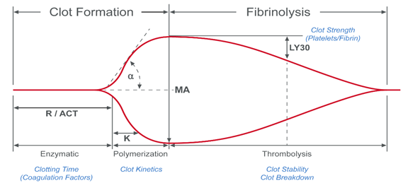
During the process of coagulation, blood experiences radical changes that cause it to lose viscosity and become more elastic.1 The resulting blood clots are able to resist deformation under shear forces, an ability called shear modulus (or modulus of rigidity). In VHA systems, the shear modulus (also known as clot stiffness) is measured either directly or indirectly to give an indication of the coagulation status of a blood sample. 

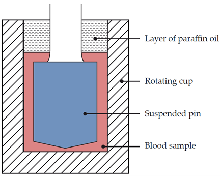
Dr Hellmut Hartert was the first to develop a mechanism to quantify the dynamics of blood clot formation, called viscoelastic haemostatic monitoring, in 1948 (Figure 1).2 The primary measurement of viscoelastic haemostatic monitoring is the observation of blood transforming from a viscous to elastic state, with VHAs assessing whole blood clotting dynamics by measuring the speed in which a blood sample can form a clot.2 By adding different reagents, investigators can determine the relative contribution of platelets, coagulation factors or fibrinogen (in cases of inadequate clotting).2 Importantly, VHAs measure the entire dynamic change of a clot and provide an overview of the haemostatic state of a blood sample, rather than just showing the presence or absence of individual coagulation factors.3

Image

Figure 1. Schematic depicting Dr Hartert's cup and pin thromboelastograph.

There are several different types of VHA with different mechanisms of action, all of which centre around detecting the level of coagulation within a whole blood sample (Table 1). 

Table 1. Overview of viscoelastic testing technology.
 DeviceMechanism of actionRotating cup with a static pin
TEG® 5000Mechanical viscoelastic testingRotating cup with a static pin
ROTEM® Delta & ROTEM® SigmaMechanical viscoelastic testingStationary cup with a rotating pin 
Sonoclot®Mechanical viscoelastic testingVertically oscillating, linear motion probe 
ClotPro®Mechanical viscoelastic testingRotating cup with a static pin
TEG® 6sResonance viscoelastic testingCartridge-based, resonance frequency device 
Quantra®Resonance viscoelastic testingCartridge-based, fully automated ultrasound device 

VHAs are currently used for the management of major bleeding as well as for guiding transfusion therapy, most commonly in the indications of:2

  • Cardiac/vascular surgery (presurgical function tests, post-operative haemostasis testing and intensive care management)

  • Trauma haemorrhage (trauma-induced coagulopathy and traumatic brain injury with associated coagulopathy) 

  • Obstetric care (management of post-partum haemorrhage)

  • Liver disease (liver transplant surgery)

  • Assisting anti-platelet therapy planning and treatment cessation

There are some limitations with current VHAs. Firstly, VHAs cannot detect platelet dysfunction alone, requiring additional assessments from PFA-100 tests.4,5  Furthermore, some VHA methodologies cannot be run on heparinised samples,5 and those that can require careful monitoring of heparin levels to avoid confounding the findings.6 Also, as with all whole-blood sampling techniques, sample interference (through improper use or even design of blood collection devices) is also present.7

Alongside the development of VHAs, haemostatic monitoring is also conducted by laboratory-based tests. These assess plasma samples for individual coagulation factors and can be time-consuming depending on laboratory availability.8 VHAs have shown advantages over laboratory haemostasis tests for:3

  • Detecting change in coagulation time

  • Detecting changes in clot lysis

  • Detecting increases in clot strength

  • Monitoring coagulation profiles over time

  • Predicting transfusion requirements

  • Rapid global assessment of clotting in whole blood

Whilst older VHA techniques have been available for several decades, new technologies are being introduced that have the potential to give clinicians a deeper understanding of their patients’ haemostasis and make viscoelastic haemostatic monitoring more readily available.2 

Available VHA devices

Within the last decade the field of viscoelastic haemostatic monitoring has expanded, with new VHA methods becoming available and with more currently under development.  Older cup and pin methods, such as thromboelastography (TEG® 5000) and rotational thromboelastometry (ROTEM® delta) have been augmented by newer cartridge systems (TEG® 6s and ROTEM®  sigma [which, whilst still utilising cartridges, is a cup and pin system]) as well as other point-of-care techniques (such as Sonoclot®, Quantra® and ClotPro®; Table 1).

What do TEG® and ROTEM® devices do?

Video: the benefits of viscoelastic haemostatic devices

Dr Seema Agarwal explaining basic principles and use of the VHA devices (TEG®5000, TEG®6s, ROTEM®Sigma) 

Thromboelastography (TEG®)

TEG® 5000

The TEG® 5000 was the first VHA device available, originally developed by Haemoscope and then acquired by Haemonetics in 2007. In the TEG® 5000 system, a 0.36 mL sample of whole blood (heated to 37 °C) is placed in a cup that rotates at 4°45’ degrees every 10 seconds. The blood sample is recalcified (if required, as in cases of non-fresh whole blood samples) and activated with reactivating agents. A pin is then placed into the cup and as a clot forms between the stationary pin and rotating cup, a force is transmitted to the pin. This force is measured as a graphical output, where the x-axis represents time (in minutes), and the y-axis represents movement of the pin (in mm; Figure 2).1

Image

Figure 2. Depiction of a TEG®5000 tracing.

The TEG® 5000 parameters include the reaction time (R), the activated clotting time (ACT), clot formation/kinetics (K), α-angle, maximum amplitude (MA), and lysis at 30 minutes (LY30; Table 2).

Table 2: TEG® parameters.
 ParameterWhat it meansWhat it measuresCoagulation phaseWhat it shows
R-value Reaction time Time of latency from start of test to initial fibrin formation Initiation phaseClotting factor availability
K-valueKineticsTime taken to achieve a certain level of clot strength (amplitude of 20 mm)Amplification phaseFibrinogen availability
α-angleSlope of line between R and K Speed of fibrin build up and cross-linking, rate of clot formationPropagation phaseFibrinogen availability
MAMaximum amplitudeOverall strength and stability of the fibrin clotFibrin formation phasePlatelets (80%) and fibrin (20%) interacting via GPIIb/IIIa
LY30Amplitude at 30 minutesPercentage decrease in amplitude at 30 minutes post-MAFibrinolysis phaseFibrin breakdown


TEG® 5000  trace patterns can help clinicians determine which clinical treatment may be most appropriate based on the shape of the traces produced (Table 3).

Table 3. A summary of TEG® trace patterns.
Process/condition

TEG® trace

Summary

Hypocoagulable Hypocoagulable TEG Trace ExampleLow clotting factors, or low platelet count/function, or low fibrinogen
HypercoagulableHypercoagulable TEG Trace ExamplePlatelet and/or enzymatic hypercoagulability
FibrinolysisPrimary fibrinolysis TEG Trace Example

Primary fibrinolysis


Secondary fibrinolysis TEG Trace ExampleSecondary fibrinolysis


TEG® 6s

The next-generation TEG® 6s device is a cartridge-based haemostasis analyser system that measures the resonance frequency of a whole blood sample exposed to frequency vibrations caused by the motion of the blood meniscus.1 It is designed to be easier to use than the TEG® 5000 device, smaller, potentially suitable for use outside of traditional clinical settings  and aims to eradicate operator-dependent variability in pipetting techniques.9
A pneumatically controlled microfluidic cartridge takes an initial blood sample and divides it across four distinct test channels (Figure 3). 

Image

Figure 3. TEG® 6s device and microfluidic TEG® 6s cartridge.

Each test channel includes a set of reagents to conduct four different assays simultaneously (Kaolin TEG®, Kaolin TEG® with Heparinase, RapidTEG® and TEG® Functional Fibrinogen). An additional PlateletMapping® cartridge also gives information on platelet function via the P2Y12 and Thromboxane A2 receptors. Rather than a rotating cup and pin, the sample is exposed to a fixed vibration resonance frequency which is measured using a light-emitting diode. The alteration of resonance is measured as the clot forms, and the collected data are converted into a graph resembling the traditional TEG® assay trace.

Rotational Thromboelastometry (ROTEM®)

ROTEM® delta

The ROTEM® delta system (depicted in Figure 4) also uses a cup and pin mechanism.1 However, the cup remains stationary whilst the pin revolves, resulting in a similar (but not identical) output to the TEG® 5000 (Figure 4). If there is no resistance to pin motion, the pin will rotate a full 4˚75’. As the clot builds up it restricts the rotation of the pin, producing the ROTEM® trace.

Image

Figure 4. Depiction of a ROTEM® delta tracing.

Whilst the ROTEM® trace looks very similar to the TEG® trace, the parameters have different names and correspond to slightly different measurements. Furthermore, the parameter numbers should not be directly compared between TEG® and ROTEM® traces as calibration settings differ between instruments and each device has different ranges for ‘normal’ levels. 

ROTEM® sigma 

The ROTEM® sigma device is, like the delta system, a cup and pin device that produces the same outputs (Figure 4). However, it is fully automated, having replaced pipetting in favour of inserting the tube of whole blood directly into the instrument via a cartridge loading system. This innovation allows it to be used as a point-of-care device at the patient’s bedside.10 As the process is fully automated, it is less time-consuming than the previous generation ROTEM® delta system.11 Furthermore, the automated sigma system requires fewer operators than the delta system, allowing for fewer operator errors to be introduced.11

ClotPro®

ClotPro® is a next-generation six-channel viscoelastometry analyser, designed for the rapid analysis of whole blood coagulation (Figure 5). 

Image

Figure 5. The ClotPro® viscoelastometry device.

ClotPro® is also a viscoelastic cup and pin device in which the pin is stationary and the cup spins.12 However, rather than relying on multiple pipetting procedures, it utilises a standard pipette which contains pre-dosed reagents that activate on contact with the whole blood sample (deposited into the cup prior to the initiation of the test run). The basic parameters are similar to those of ROTEM® delta, though ClotPro® has additional specialised testing capabilities, listed as specific assays (Table 4).1 

Table 4: Specialised ClotPro® assays.

 AssayActivator/inhibitorDescription
EX-testTissue factor (TF)Assess the extrinsic coagulation pathway its interaction with platelets in citrated blood
IN-testFactor VIIIAssess the intrinsic coagulation pathway and the effect of heparin
HI-testFactor VIIIThe IN-test but with heparin inhibition
RVV-testDirect oral anticoagulantsDetects factor Xa antagonists in citrated blood
ECA-testDirect thrombin inhibitorsSensitive to and detects direct thrombin inhibitors such as dabigatran
NA-test-Non-activated test
FIB-testCytochalasin D and a synthetic GPIIb/IIIa antagonistExamines levels of fibrinogen and fibrin polymerisation in citrated blood
AP-testAprotininFibrinolysis is inhibited in vitro
TPA-testRecombinant tissue plasminogen activator Permits the in vitro detection of anti-fibrinolytic agents in citrated blood


Other devices (Quantra® and Sonoclot®)

Quantra®

The Quantra® device (Figure 6) uses sonic estimation of elasticity via resonance (SEER), also known as sonorheometry, to estimate the firmness of a sample within a test chamber by measuring clot time and stiffness with ultrasound.1 It is a cartridge-based, fully automated, four-channel device that can organise its test results into three different views: trend data, clot stiffness curves and dial results. The results are displayed on a digital screen.

Image

Figure 6. The Quantra® device.

The Quantra QPlus® cartridge is inserted directly into the machine and carries out all assays:

  • Channel 1 assesses clot coagulation time with kaolin
  • Channel 2 adds heparinase I enzyme and measures the relative coagulation time 
  • Channel 3 is activated by thromboplastin and measures clot stiffness due to fibrin polymerisation and platelet adhesion/aggregation 
  • Channel 4 adds abciximab to evaluate fibrinogen contribution 
  • Two additional parameters are calculated: 
    • Clot time ratio 
    • Platelet contribution 

SonoClot®

The SonoClot® device is a vertical oscillating probe system that vibrates inside the whole blood sample. An empty plastic probe is attached to the head of the transducer and submerged into the sample that contains different coagulation activators or inhibitors, depending on the assay.2 The probe oscillation transmits a force to the transducer, which emits a signal proportional to the deflection of the tube, producing an oscillatory pattern directly linked to the viscoelastic properties of the sample.1 The results are shown as a graph (Figure 7); on the x-axis is time and on the y-axis movement (in mm) is recorded. 

Image

Figure 7. Depiction of a SonoClot® tracing. 

Related Content

References

References

  1. Volod O, et al. Viscoelastic Hemostatic Assays: A Primer on Legacy and New Generation Devices. J Clin Med 2022; 11(3):860.
  2. Hartmann J, et al. Viscoelastic Hemostatic Assays: Moving from the Laboratory to the Site of Care-A Review of Established and Emerging Technologies. Diagnostics (Basel) 2020; 10(2):118.
  3. Peng HT, et al. A Comparative Study of Viscoelastic Hemostatic Assays and Conventional Coagulation Tests in Trauma Patients Receiving Fibrinogen Concentrate. Clin Chim Acta 2019; 495: 253─62.
  4. Paniccia R, et al. Platelet Function Tests: A Comparative Review. Vasc Health Risk Manag 2015; 11: 133─48.
  5. Ranucci M, et al. Sensitivity of Viscoelastic Tests to Platelet Function. J Clin Med 2020; 9(1):189.
  6. Dias JD, et al. Cartridge-Based Thromboelastography Can Be Used to Monitor and Quantify the Activity of Unfractionated and Low-Molecular-Weight Heparins. TH Open 2019; 3(3): e295─e305.
  7.  Bowen RA, et al. Interferences From Blood Collection Tube Components on Clinical Chemistry Assays. Biochem Med (Zagreb) 2014; 24(1): 31─44.
  8. Rali AS, et al. Viscoelastic Haemostatic Assays in Cardiovascular Critical Care. Card Fail Rev 2020; 7: e01.
  9. TEG 6s Hemostasis Analyzer | Haemonetics®. https://teg.haemonetics.com/en-gb/teg-6s-hemostasis-analyzer (accessed 02/02/2023).
  10. Gillissen A, et al. Comparison of Thromboelastometry by ROTEM(®) Delta and ROTEM(®) Sigma in Women With Postpartum Haemorrhage. Scand J Clin Lab Invest 2019; 79(1─2): 32─8.
  11. Schenk B, et al. A Comparison of the New ROTEM(®) Sigma With Its Predecessor, the ROTEM Delta. Anaesthesia 2019; 74(3): 348─56.
  12. Leyser H. Devices and Methods for Measuring Viscoelastic Changes of a Sample. Patent No. WO 2018/137766 Al. 2018. https://patentimages.storage.googleapis.com/08/d3/f3/f31384ed000c19/WO2018137766A1.pdf (accessed 03/02/2023).