This educational website was funded by an educational grant provided by Haemonetics. Additionally, certain content references and imagery on this website were also provided by Haemonetics.

Clotting

  • Clotting involves three stages: initiation, amplification and propagation
  • Activated platelets form an initial plug (primary haemostasis), which is then stabilised within a mesh of fibrin, forming a clot (secondary haemostasis)
  • Fibrinolysis is the process of breaking down a fibrin clot and is mediated by plasmin, which is converted from plasminogen
  • Coagulation and fibrinolysis can be assessed through laboratory tests
Image
clotting blood cells

Introduction 

In the contemporary view of haemostasis, clotting includes three distinct phases: initiation, amplification and propagation (Figure 1)1, 2.

- Initiation involves the exposure of tissue factor to coagulation factors, with subsequent activation of other factors and the generation of a small amount of thrombin, which converts fibrinogen to fibrin.

- In the amplification phase, coagulation factor production is increased and thrombin production is accelerated. This also promotes maximal platelet activation1, 2.

- Finally, during propagation, activated platelets are recruited to the site of injury and there is a burst of thrombin production. This leads to the conversion of fibrinogen to fibrin and the production of a stable fibrin clot1-3.

Image

Figure 1: The three stages of coagulation.2

Platelets and Fibrin

Platelets are small, flat, disc-shaped blood cells that circulate in the blood and help to form blood clots.4 In the event of vascular injury, platelets are activated by adhesion to von Willebrand factor or collagen, and by circulating soluble platelet-activating agonists. The activation causes them to change shape, release further platelet activating agonists, including ADP and thromboxane A2,5,6 and aggregate to form a primary platelet plug (primary haemostasis).1 Further platelets are activated by these circulating agonists, or by fibrinogen binding to the GpIIb/IIIa receptors. Fibrin monomers polymerise into a fibrous gel,4 and stabilise and grow within this mesh of fibrin and aggregated platelets, forming a clot (secondary haemostasis).1

Fibrinolysis

Fibrinolysis is the process of removing a fibrin clot once the injured blood vessel has healed.7 The principal mediator of this is plasmin, which breaks down the fibrin polymer to produce soluble fibrin degradation products.2,7–9Plasmin is produced from circulating plasminogen, which is synthesised in the liver.2,8,9 The conversion of plasminogen to plasmin is facilitated by two types of plasminogen activators, released from endothelial cells (Figure 2).2,8

As the presence of plasmin increases plasminogen activator activity, plasminogen therefore exerts positive feedback on its own activation and ensures the fibrin clot is removed appropriately.7 To avoid excessive degradation of fibrin and to maintain balanced haemostasis, plasminogen activator inhibitors also regulate this process by forming inactivating complexes with the plasminogen activators to prevent inappropriate plasmin generation.2,10

Image

Figure 2. Overview of the fibrinolytic system.9

Conventional laboratory tests for clotting

Laboratory tests and assessments are able to specifically identify the levels of individual components of the coagulation pathway, which can help diagnose the cause of patient bleeding or clotting (Table 1). They use plasma samples, as opposed to whole blood, with levels of coagulation components assessed separately through individual tests.11

Table 1. Standard coagulation tests.
 TestApproximate normal range12Description
Platelet count13,14150–350 x 109 cells/L

There are four main methods of counting the number of platelets in the blood: 

  1. manual counting using phase contrast microscopy;

  2. impedance analysis;

  3. optical light scatter/fluorescence analysis;
  4. immunoplatelet counting by flow cytometry.
Activated partial thromboplastin time (aPTT)1425–35 seconds (ratio to normal: 0.8–1.2)A measure of the time necessary to generate fibrin via the intrinsic pathway of the coagulation cascade. Assesses the levels of factors XII, IX, VIIII, X, V, prothrombin, and fibrinogen.
Prothrombin time (PT)1511–13 seconds (ratio to normal: 0.8–1.2)

A measure of the time necessary to generate fibrin via the extrinsic pathway of the coagulation cascade (i.e., levels of factors VII, V, X, prothrombin, and fibrinogen).

The international normalised ratio (INR) can be derived from the PT for patients receiving vitamin K antagonists as a way to harmonise results across different thromboplastins16.

Thrombin time1418.5–24 secondsA measure of the rate of fibrinogen conversion to fibrin in the presence of thrombin. Identifies changes in the levels of fibrinogen and if there are any inhibitors to fibrinogen activation.
Fibrinogen assay17-191.5–3.5 g/L

The level of fibrinogen is assessed based on clotting time of diluted plasma exposed to high concentrations of thrombin, which is then compared against a standard calibration curve. There are two main methods for assessing fibrinogen levels:

  1. Clauss assay;

  2. PT-derived (PT-Fg) tests

Activated clotting time20VariableA commonly used assay for monitoring anticoagulation during extracorporeal life support. The assay uses whole blood, so also includes measurement of platelets and phospholipids.

Assessing fibrinolysis

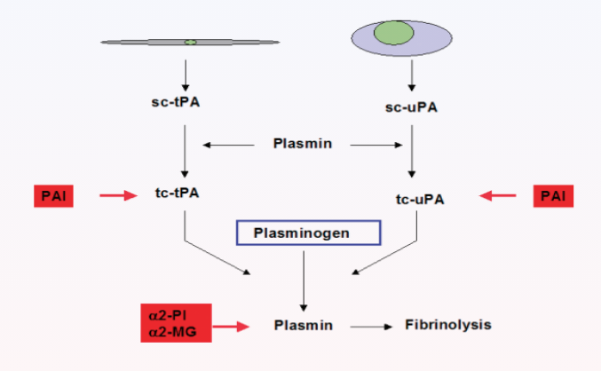
Fibrinolysis is difficult to measure directly using conventional laboratory tests.7 Standardised methods to detect abnormalities in fibrinolysis are not as simple or widely available as those used to measure blood clotting.21–23

Assessment of euglobulin clot lysis time can be used as a measure of fibrinolysis. Alternatively, antigen tests for plasma proteins involved in fibrinolysis, such as tissue plasminogen activator (tPA), plasminogen activator inhibitor 1 (PAI-1) and thrombin-activatable fibrinolysis inhibitor (TAFI) can be assessed.21–23 Fibrin degradation products, such as D-dimers, are another parameter that can be measured. 

D-dimers are late-stage fragments released when fibrin, cross-linked by factor XIII, is degraded by plasmon.9  Circulating D-dimer levels are therefore a measure of fibrin breakdown and ongoing fibrinolysis.9,12  In a clinical setting, D-dimer tests are restricted to the exclusion of thrombosis, due to their low specificity.21,22

Limitations of laboratory tests for haemostasis

The process of obtaining laboratory tests can be time consuming and often takes too long to be able to provide real-time guidance in a clinical setting.11,23 Haemostasis is a complex process in vivo, and consequently individual laboratory tests only give a narrow view of the clinical picture. They are often performed on plasma samples without platelets and other blood cells, and they do not always differentiate between the various pathological mechanisms that may be contributing to a specific clinical situation.11 Some tests are also susceptible to interference from particular serum constituents or antibodies,8,21 and some comorbidities, inflammatory mediators or commonly administered treatments can also adversely affect the sensitivity of certain assays.7,11

Related Content

References

  1. Monroe DM, et al. The Coagulation Cascade in Cirrhosis. Clin Liver Dis. Feb 2009;13(1):1-9. doi:10.1016/j.cld.2008.09.014
  2. Adams RL, et al. Review Article: Coagulation Cascade and Therapeutics Update: Relevance to Nephrology. Part 1: Overview of Coagulation, Thrombophilias and History of Anticoagulants. Nephrology. Aug 2009;14(5):462-70. doi:10.1111/j.1440-1797.2009.01128.x
  3. Gailani D, et al. Intrinsic Pathway of Coagulation and Arterial Thrombosis. Arterioscler Thromb Vasc Biol. Dec 2007;27(12):2507-13. doi:10.1161/atvbaha.107.155952
  4. Fogelson AL, et al. Fluid Mechanics of Blood Clot Formation. Annu Rev Fluid Mech. Jan 1 2015;47:377-403. doi:10.1146/annurev-fluid-010814-014513
  5. Sang Y, et al. Interplay between Platelets and Coagulation. Blood Rev. Mar 2021;46:100733. doi:10.1016/j.blre.2020.100733
  6. Li Z, et al. Signaling During Platelet Adhesion and Activation. Arterioscler Thromb Vasc Biol. Dec 2010;30(12):2341-9. doi:10.1161/ATVBAHA.110.207522
  7. Chapin JC, et al. Fibrinolysis and the Control of Blood Coagulation. Blood Rev. Jan 2015;29(1):17-24. doi:10.1016/j.blre.2014.09.003
  8.  Raber MN. Coagulation Tests. In: Walker HK, Hall WD, Hurst JW, eds. Clinical Methods: The History, Physical, and Laboratory Examinations. 3rd ed. Butterworths; 1990:chap 157.
  9. Cesarman-Maus G, et al. Molecular Mechanisms of Fibrinolysis. Br J Haematol. May 2005;129(3):307-21. doi:10.1111/j.1365-2141.2005.05444.x
  10. Longstaff C, et al. Basic Mechanisms and Regulation of Fibrinolysis. J Thromb Haemost. Jun 2015;13 Suppl 1:S98-105. doi:10.1111/jth.12935
  11. Fowler A, et al. Laboratory Monitoring of Haemostasis. Anaesthesia. Jan 2015;70 Suppl 1:68-72, e24. doi:10.1111/anae.12919
  12. Merck & Co. Inc. Msd Manual. Blood Tests: Normal Values. https://www.msdmanuals.com/en-gb/professional/resources/normal-laboratory-values/blood-tests-normal-values
  13.  National Heart, Lung, and Blood Institute. Thrombocytopenia. https://www.nhlbi.nih.gov/health/thrombocytopenia
  14. Briggs C, et al. Continuing Developments with the Automated Platelet Count. Int J Lab Hematol. Apr 2007;29(2):77-91. doi:10.1111/j.1751-553X.2007.00909.x
  15. Tripathi MM, et al. Clinical Evaluation of Whole Blood Prothrombin Time (Pt) and International Normalized Ratio (Inr) Using a Laser Speckle Rheology Sensor. Sci Rep. Aug 23 2017;7(1):9169. doi:10.1038/s41598-017-08693-5
  16. Tripodi A, et al. How to Report Results of Prothrombin and Activated Partial Thromboplastin Times. Clin Chem Lab Med. Feb 2016;54(2):215-22. doi:10.1515/cclm-2015-0657
  17. Miesbach W, et al. Comparison of the Fibrinogen Clauss Assay and the Fibrinogen Pt Derived Method in Patients with Dysfibrinogenemia. Thromb Res. Dec 2010;126(6):e428-33. doi:10.1016/j.thromres.2010.09.004
  18.  Mackie IJ, et al. Guidelines on Fibrinogen Assays. Br J Haematol. May 2003;121(3):396-404. doi:10.1046/j.1365-2141.2003.04256.x
  19. Stang LJ, et al. Fibrinogen. Methods Mol Biol. 2013;992:181-92. doi:10.1007/978-1-62703-339-8_14
  20. Horton S, et al. Activated Clotting Time (Act). Methods Mol Biol. 2013;992:155-67. doi:10.1007/978-1-62703-339-8_12
  21. Longstaff C. Measuring Fibrinolysis: From Research to Routine Diagnostic Assays. J Thromb Haemost. Apr 2018;16(4):652-662. doi:10.1111/jth.13957
  22.  Longstaff C. Measuring Fibrinolysis. Hamostaseologie. Feb 2021;41(1):69-75. doi:10.1055/a-1325-0268
  23. Dias JD, et al. Global Coagulation Assays to Measure in Vitro Fibrinolysis. Thrombosis Update. 2021/08/01/ 2021;4:100052. doi:https://doi.org/10.1016/j.tru.2021.100052